The alimentary canal is a long hollow tube which runs from the mouth to the anus.
Together with several other organs, including the liver and the pancreas, it makes up the digestive system.
The total length of the human alimentary canal is between 5 and 6 m, from anus to mouth.
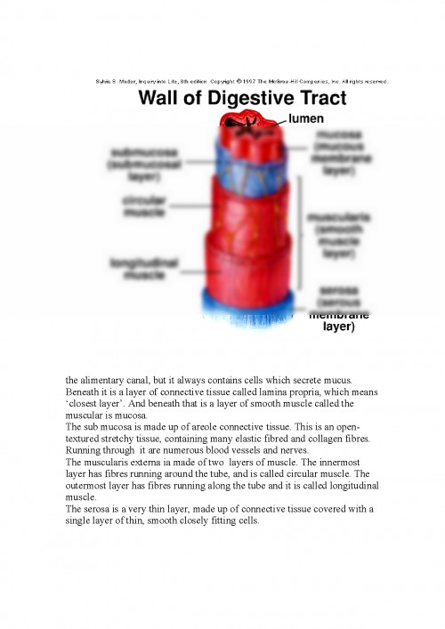
To fit this considerable length into body, parts of the canal are folded and coiled inside the abdomen. The mucus is a substance secreted along the tube by cells lining its walls. Mucus helps food to slide through the canal without doing too much damage to the lining. It also forms a protective covering which keeps the digestive juices, which are inside the lumen of the canal, from coming into contact with the living cells of the walls. Along the whole length of the alimentary canal there are muscles in the walls. These produce waves, of Contraction and relaxation called peristaltic waves, which move food along the alimentary canal and help to mix the contents. Each region of the alimentary canal has it own function and different structure. There are 4 basic layers in the wall of the alimentary canal.
Working from the inside these are: a) the mucosa b) the submucosa c) the muscularis externa d) the serosa. Many of this names came from Latin origin.
The mucosa is made up of 3 layers. The innermost layer is the epithelium. The structure of the epithelium varies in different parts of Beneath it is a layer of connective tissue called lamina propria, which means closest layer. And beneath that is a layer of smooth muscle called the muscular is mucosa.
The sub mucosa is made up of areole connective tissue. This is an open-textured stretchy tissue, containing many elastic fibred and collagen fibres. Running through it are numerous blood vessels and nerves.
The muscularis externa ia made of two layers of muscle.
The innermost layer has fibres running around the tube, and is called circular muscle.
The outermost layer has fibres running along the tube and it is called longitudinal muscle.
The serosa is a very thin layer, made up of connective tissue covered with a single layer of thin, smooth closely fitting cells.
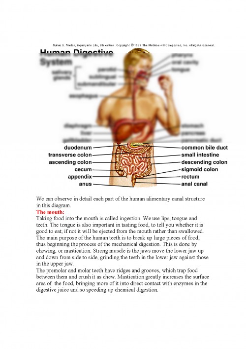
We can observe in detail each part of the human alimentary canal structure in this diagram.
The mouth: Taking food into the mouth is called ingestion. We use lips, tongue and teeth. The tongue is also important in tasting food, to tell you whether it is good to eat; if not it will be ejected from the mouth rather than swallowed.
The main purpose of the human teeth is to break up large pieces of food, thus beginning the process of the mechanical digestion.
This is done by chewing, or mastication. Strong muscle is the jaws move the lower jaw up and down from side to side, grinding the teeth in the lower jaw against those in the upper jaw.
The premolar and molar teeth have ridges and grooves, which trap food between them and crush it as chew. Mastication greatly increases the surface area of the food, bringing more of it into direct ...
Pentru a descărca acest document,
trebuie să te autentifici in contul tău.